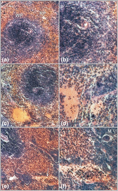
Figure 5.

Mouse spleen after mobilization with G‐CSF plus Cy. The domination of the red pulp (RP) over the white pulp (WP) of spleen after 2 days (a,b) of mobilization, and dilated parenchymal vessels filled with erythrocytes and leukocytes on the 4th day (c,d). The parenchymal vessels filled with a higher number of leukocytes (arrow) on the 6th day of mobilization (e,f). Megakaryocytes (M) are visible. Haematoxylin & eosin (a,c,e) magnification × 330, (b,d,f) magnification × 670 (magnifications at time of photography).
