
Figure 4.
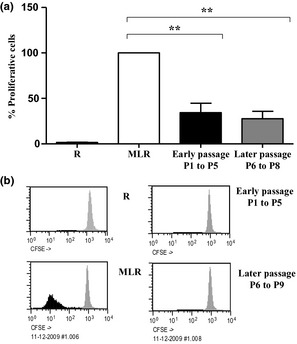
hMSC‐mediated T cell inhibition. (a) hMSC inhibition of MLR. Lymphocyte proliferation was assessed using MLR in absence (MLR) or presence of 10% hMSCs/well, administered at early (P1 to P5) or late (P6 to P10) passage. Results expressed as mean ± sd for independent experiments using 9 hMSC cultures (P < 0.01); (b) Representative assay showing MLR in absence (MLR) or presence of 10% MSC/well at early (P1 to P5) or late (P6 to P9) passages. Black represents proliferative cells and grey represents non‐proliferative cells. R ‐ responders without stimulators.