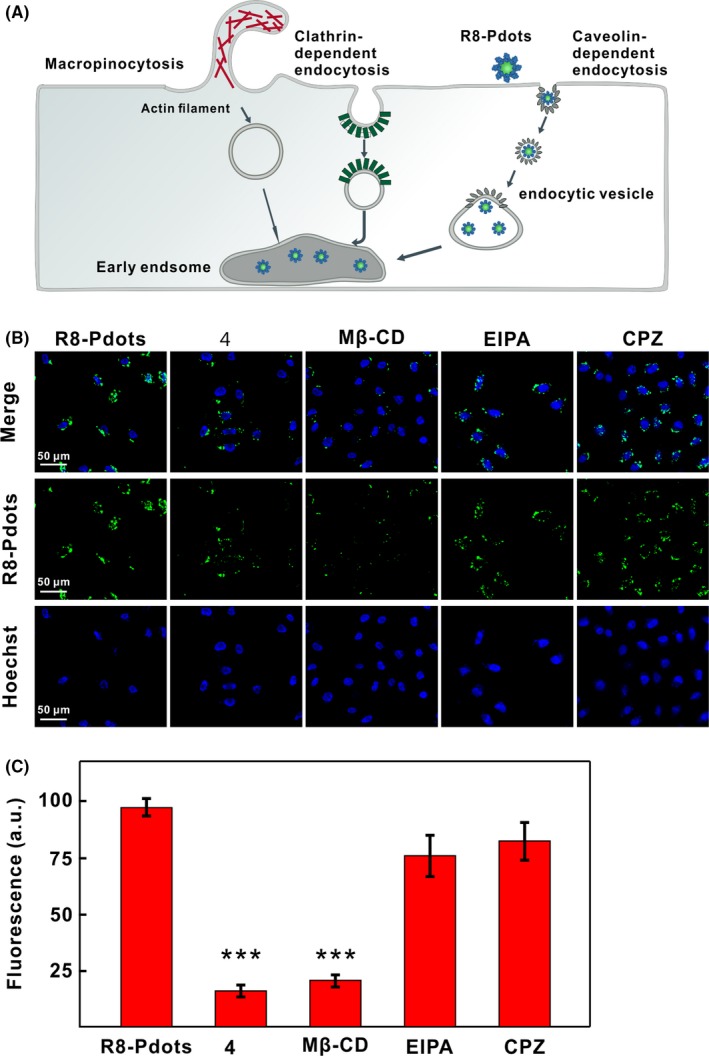
Figure 3.

Endocytic pathway of R8‐Pdots. A, Schematic illustration of various endocytotic routes and internalization of R8‐Pdots by HeLa cells. B, Confocal images of HeLa cells treated with indicated pharmacological inhibitors. Cells were pre‐treated with low temperature (4°C), mβ‐CD, EIPA or CPZ for 30 minutes, followed by incubation with 5 μg/mL R8‐Pdots for another 60 minutes. C, Intracellular fluorescent signals of cells treated in the presence of different inhibitors were quantified using the ImageJ software (20 cells analysed)
