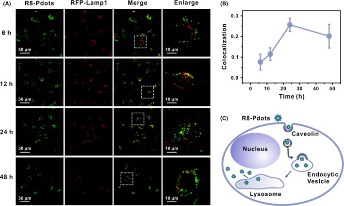
Figure 5.

R8 modification facilitates endosomal escape of Pdots. HeLa cells expressing RFP‐Lamp1 (red) were incubated with 5 μg/mL R8‐Pdots (green) and imaged by confocal microscope at indicated time points. Right panel shows magnified images of the square region in the left panel. B, Colocalization ratio of R8‐Pdots with LAMP1 was quantified using the ImageJ software (20 cells analysed). C, Schematic illustration of intracellular distribution and transportation of R8‐Pdots
