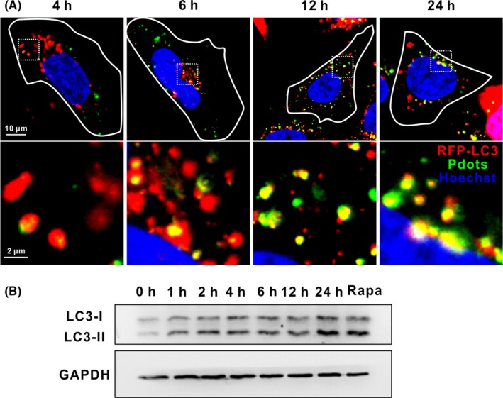
Figure 6.

R8‐Pdots induce autophagy. HeLa cells expressing RFP‐LC3 (red) were incubated with 20 μg/mL Pdots (green) and imaged by confocal microscope at indicated time points. Lower panel shows magnified images of the square region in the upper panel. B, HeLa cells were incubated with 5 μg/mL R8‐Pdots for indicated time. Cells treated with 100 μM rapamycin were included as a positive control. Protein levels of LC3 were analysed by Western blotting. GAPDH was included as loading control
