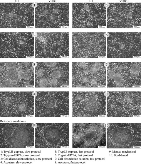
Figure 2.

Phase contrast microscopy of H1 and VUB01 hESC colonies under the different experimental conditions. Morphologically, there is no difference visible in the first passage between the colonies formed by H1 cell line and VUB01 cell line, under the same experimental conditions. In general, colonies formed with TrypLE™ Express and Trypsin‐EDTA are the smallest. Those formed by Cell Dissociation Solution are comparable to the reference conditions and those formed with Accutase are of an intermediate size.
