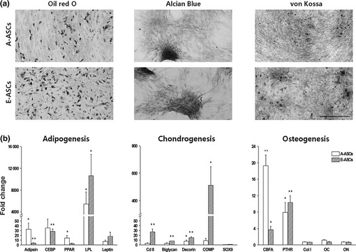
Figure 3.

In vitro differentiation of ASCs into mesodermal lineages. (a) A‐ASCs and E‐ASCs were cultivated in adipogenic, chondrogenic or osteogenic differentiation medium. After culture, adipogenic differentiation of cells was assessed by intracellular accumulation of neutral lipids following staining with Oil red O solution. Chondrogenic differentiation was assessed by staining with Alcian blue. Formation of a mineralized matrix after osteogenic differentiation was assessed by von Kossa staining. Scale bar: 500 μm. (b) Quantification of mesodermal lineage‐specific gene expression. Total RNA was extracted from differentiated cells, and was analysed by qRT‐PCR. Gene expression levels were normalized to GAPDH expression to yield 2Ct values, and were calculated relatively to the controls.
