Figure 3.

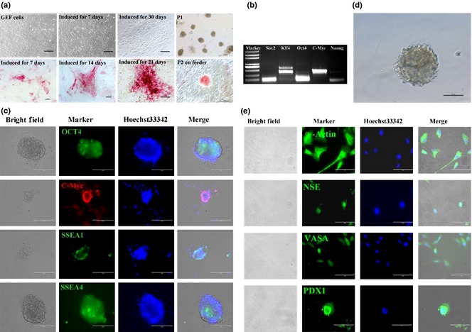
Characterization of dairy goat iPSCs. (a) Induced goat iPSC‐like colonies were positive for AP staining; (b) RT‐PCR analysis showed that goat iPSC‐like cells were positive for SOX2, KLF4, OCT4, C‐MYC and NANOG; (c) immunofluorescence analysis of dairy goat iPSCs at passage 2; pluripotent relative markers (OCT4, C‐MYC, SSEA1, SSEA4) were expressed. Nuclei were counterstained with Hoechst 33342. (d) Goat iPSCs formed embryoid bodies (EBs). (e) Immunofluorescent staining showed that the EBs derived from goat iPSCs differentiated into three germ layer cell types, including the NSE, α‐ACTIN, PDX1 and VASA‐positive cells. Goat iPSCs differentiated into EBs.
