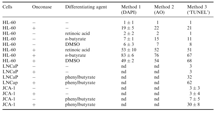
Table 1.
Percent of apoptotic cells in HL‐60, LNCaP and JC‐1 cultures untreated and treated with 0.42 µM Onc in the absence or presence of differentiating agents. Apoptotic cells were identified by two methods utilizing different DNA fluorochromes, DAPI and acridine orange (AO), and the third one, based on the presence of DNA strand breaks ( Gorczyca et al. 1992 ) as shown in 2, 3, 4. Their frequency was estimated by flow cytometry, as described in Materials and Methods. nd, not done