
Figure 5.
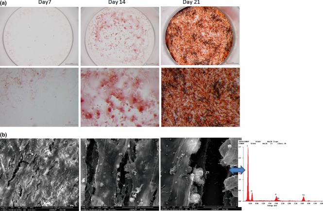
Matrix mineralization. (a) Alizarin red histochemical staining performed in co‐cultured cells, at 7, 14 and 21 days. (b) SEM images of HMSC and HDMEC co‐cultured after 21 days. Mineralized globular structures were identified in close association with cell layers, and EDS spectrum showed the presence of Ca and P peaks.