
Figure 1.
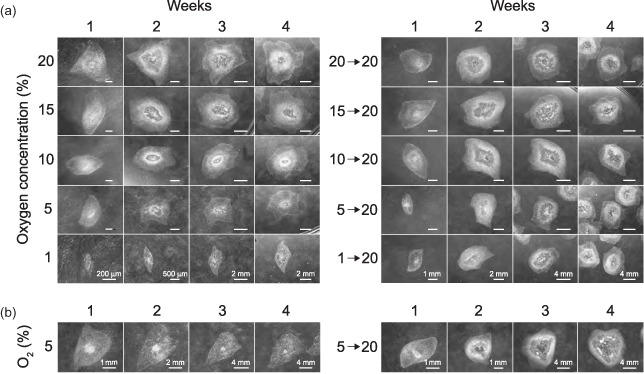
The effect of short‐ and long‐term hypoxic exposure and re‐oxygenation on the morphology of hESC colonies. (a) The CLS1 line was sub‐cultured by mechanical dissection and grown for a period of 4 weeks in five different concentrations of atmospheric oxygen, covering the range from 1% to 20%. After the initial treatment, colonies from all conditions were grown at oxygen concentration of ambient air for additional 4 weeks. (b) The CLS1‐LT line has been continuously maintained in hypoxic conditions corresponding to 5% oxygen for over 18 months. The development of colony morphology between regular passaging intervals is shown over the course of 4 weeks. The response to re‐oxygenation corresponding to the oxygen concentration of ambient air was followed during the course of additional 4 weeks. For each gaseous condition, the development of a representative colony is shown. The images were taken with a maximum zoom, given the size of a particular colony, and stereo microscope illumination settings were adjusted to a dark field observation. Image scalings at the bottom line images apply to all images within the specific week. hESC, human embryonic stem cell.