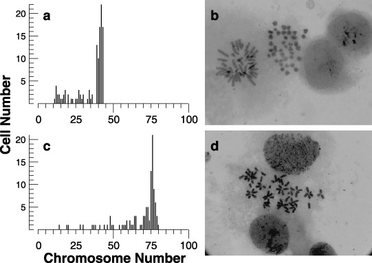
Figure 4.

Histogram of chromosome number (a and c) and photomicrographs (b and d) of 2nH1 (a and b) and 4nH1 cells (c and d). Exponentially growing 2nH1 and 4nH1 cells were exposed to 270 nm DC for 3 h. Giemsa‐stained chromosomes of about 100 cells were enumerated from enlarged photographs.
