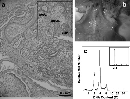
Figure 7.

A histological section (a), an abdominal photograph (b) and a DNA histogram (c) of solid tumours formed by 4nH1 cells after intraperitoneal injection into a normal C3H/He mouse. 4nH1 cells growing exponentially were intraperitoneally injected. Solid tumours were formed in several organs (b). The histological section of the tumour showed a heterogeneous conformation of cells (a). In the insert of a, endodermal, mesodermal and ectodermal cells are indicated as endo., meso. and ecto., respectively. The tumour on a liver showed DNA histograms with 2C, 4C and 8C peaks, suggesting that it is tetraploid (c). The insert of DNA histogram c is of normal tissue around the tumour. In the DNA histograms, the abscissa represents DNA content (C, complement).
