
Figure 4.
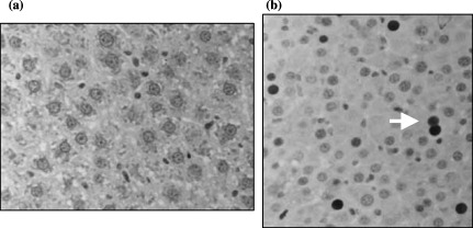
(a) Anti‐PCNA immunohistochemical staining of a control Gunn rat liver treated with RS (40×). (b) Anti‐PCNA immunohistochemical staining of the host liver of a Gunn rat transplanted with foetal hepatocytes, treated with RS and T3, 30 days after transplantation (40×) (arrow: positively stained nucleus). Each picture shows a part of a field. As explained in Material and Methods the LI is determined from 10 fields randomly chosen in each slide.