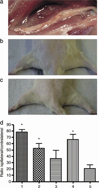
Figure 1.

Macroscopic examination six months after nerve grafting. (a) Anatomic microdissection of the regenerated sciatic nerve. A 20 mm gap in the nerve had been interposed with an isogenic vein filled with spider silk fibres. The graft has maintained its original volume, is well integrated into the host tissue and well vascularized. (b) Comparison between the ipsilateral and contralateral gastrocnemius muscles in an animal grafted with veins and matrigel. On the left, the control is shown, compared to which the muscle on the right has atrophied following denervation. (c) Comparison between the ipsilateral and contralateral gastrocnemius in an animal grafted with veins and spider silk. On the left, the control is displayed, compared to which the muscle on the right has completely retained its mass. (d) Muscle to weight ratio of the ipsilateral and contralateral gastrocnemius muscles. Experimental groups are indicated according to Table 1. Data represent means ± SEM (n= 5). Statistical analysis was performed with paired student's t‐test and adjusted according to Bonferroni. Stars indicate significant differences (P < 0.05).
