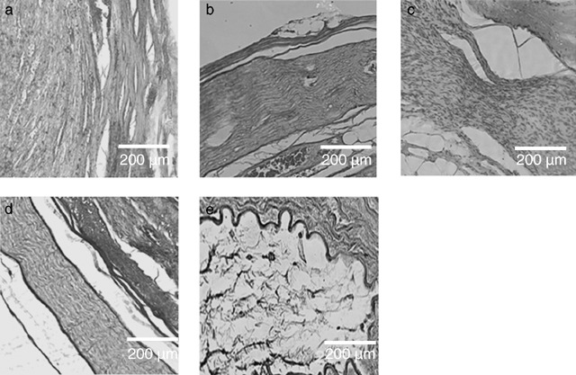
Figure 2.

Comparative examination of the five experimental groups. Hamatoxylin‐eosin staining of paraffin‐embedded distal parts of the analyzed nerve grafts, harvested six months after implantation. Longitudinal sections were analyzed under a light microscope. The original magnification was 100×. (a) Isogenic nerve graft. Neural tissue is regularly patterned along the longitudinal axis. (b) Artificial nerve conduit consisting of vein and spider silk. The neural tissue is regularly patterned. Note the epineural and perineural sheath at the margin of the construct. (c) Artificial nerve conduit consisting of vein, spider silk and Schwann cells. (d) Artificial nerve conduit consisting of vein, spider silk, Schwann cells and matrigel. Axons are aligned along the longitudinal axis and epineural and perineural sheaths are visible. (e) Artificial nerve conduit consisting of vein and matrigel. Axonal regeneration is not visible, and no cells can be detected inside the conduit.
