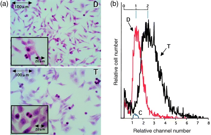
Figure 5.

Micrographs (a) and volume distribution (b) of diploid (D) and triploid (T) V79 cells. Exponentially growing diploid and triploid V79 cells were prepared for measurements. In a, the cells were stained with HE. In b, longitudinal lines (blue) were drawn to facilitate understanding. C is of standard spheres 9.8 µm in diameter.
