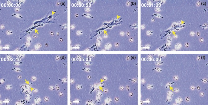
Figure 2.

HCs engaged and lifted together with mMSCs in primary bone marrow adherent culture. (a–f) Representative sequential frames from time‐lapse video microscopic recording, showing one HC engaged on top of mMSCs, at the beginning of the recording, which lifted together with mMSCs by trypsin digestion within 6 min (arrowhead). HCs adjacent to but not overlapped with mMSCs (arrow) were not lifted. Scale bars represent 50 μm.
