Figure 5.

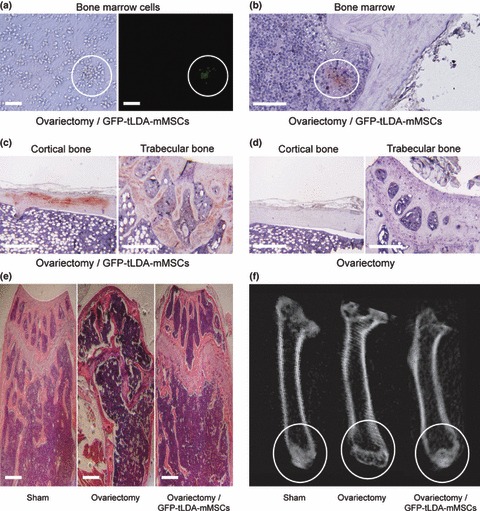
Repair of ovariectomy‐induced bone loss in mice after injection of tLDA‐mMSCs. Mice undergoing osteoporotic injury were injected intravenously with GFP‐tLDA‐mMSCs. (a) Phase‐contrast (left) and fluorescence (right) images of bone marrow cells isolated from osteoporotic mice 2 months after injection of GFP‐tLDA‐mMSCs. (b–c) GFP positive signal was found in bone marrow (b), cortical bone (left) and trabecular bone (right) (c) by IHC staining. (d) Control tissue (left: cortical bone; right: trabecular bone) from osteoporotic mice injected with PBS showed no GFP staining. (e) Histological section of femurs taken from sham‐operated mice (left), OVX mice (middle) and OVX mice receiving GFP‐tLDA‐mMSCs (right). Scale bars represent 50 μm. (f) Images of μCT scans of femurs taken from sham‐operated mice (left), OVX mice (middle) and OVX mice receiving GFP‐tLDA‐mMSCs (right) at 10 μm resolution.
