
Figure 1.
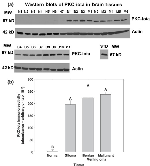
PKC‐iota is present in benign and malignant meningiomas, gliomas but not normal brain tissue. (a) Human autopsy‐derived normal brain tissue (N1, frontal lobe; N2, cortex; N3 and N4, unspecified brain; N5, cortex; N6, cerebellum), benign tumour tissue (B1, B4, B7, B9 and B10, [WHO grade 1] meningothelial meningioma; B5 and B8, meningioma; B6, fibroblastic meningioma; B2, B3 and B11, fibrous meningioma [WHO grade 1], and malignant tumour tissue; M1 and M2, [WHO grade IV] glioblastoma multiforme; M3, right frontal lobe meningioma; M4, atypical meningioma [WHO grade II]; M5, Astrocytoma [WHO grade IV]; M6, anaplastic meningioma [WHO grade III]). Specimens were obtained from the Cooperative Human Tissue Network. (b) Immunoblots from 12 normal brain specimens, 3 gliomas, 15 benign meningiomas and 3 malignant meningiomas were quantified, and mean plus and minus SE value is presented for each tissue type. Treatments indicated by the same letter do not differ, according to Tukey's honestly significant difference test (P = 0.001).