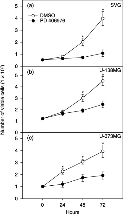
Figure 6.

Cytocidal effects of PD 406976 on (a) SVG, (b) U‐138MG and (c) U‐373MG cells. Cells were plated on 75 cm2 flasks at a density of 1.0 × 10⁶ cells/flask. Twenty‐four hours post‐plating, cells were incubated with either DMSO (vehicle; control) or PD 406976 (2 µm, dissolved in DMSO). Following a 3‐day incubation with either DMSO or PD 406976, the number of viable cells were quantified by trypan blue dye exclusion assay. Results are means ± SEM of three independent experiments. (○) Control (DMSO‐treated) cells; (•) solid symbols represent cells treated with PD 406976 (2 µm).
