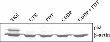
Figure 6.

Immunoblotting analyses of p53 protein in control and treated KYSE‐510 cells. Twenty micrograms of protein extracts from control cells and cells incubated 24 h in fresh medium after the end of individual (PDT; CDDP 1 μm) or combined (PDT + CDDP 1 μm) treatments, were subjected to 12% SDS–PAGE. TK6 cells irradiated with 5Gy of γ‐rays were used as positive control for p53 detection.
