
Figure 8.
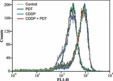
Flow cytometric analyses of green (FL‐1) fluorescence of the potential sensitive probe JC‐1 in KYSE‐510 cells. The increased fluorescence from JC‐1 monomers indicated the dissipation of the mitochondrial membrane potential. Analyses were carried out at 24 h after the end of treatments with PDT alone, CDDP alone (1 μm) and their combination.