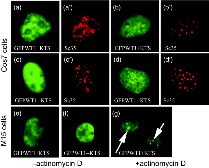
Figure 4.

In transcriptionally quiescent cells, WT1 displays isoform‐specific nuclear distributions. Cos7 cells were transfected with GFPWT1 isoforms and incubated for 48 h before actinomycin D treatment was done to render them transcriptionally quiescent. The nuclear distribution of each GFPWT1 isoform and the splicing factor Sc35 were detected using GFP fluorescence (green) and texas‐red conjugated antibody to Sc35 (Red). GFPWT1+KTS and Sc35 before actinomycin D treatment (a and a′), GFPWT1‐KTS and Sc35 before actinomycin D treatment, (c and c′). GFPWT1+KTS and Sc35 after treatment (b and b′). GFPWT1−KTS and Sc35 after treatment (d and d′). 24 h after expression in M15 cells, the discrete nuclear distribution of GFPWT1+KTS (e) and GFPWT1‐KTS (f) were indistinguishable. However, following actinomycin D treatment, the +KTS isoform was again seen to re‐locate to the nucleolus (g, arrows).
