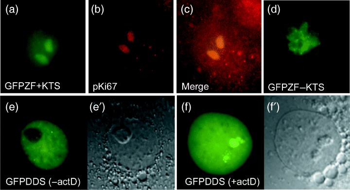
Figure 6.

The zinc finger region determines sub‐nuclear distribution of WT1 isoforms. Nuclear distribution of fusions between GFP and only the WT1 zinc finger regions (GFPZF) in live Cos7 cells 48 hpt (a and d). Only nucleolar accumulation was seen in all cells expressing GFPZF+KTS (a). The nucleolar localization was confirmed using texas‐red conjugated antibody detection of B23 (b and merge c). Nuclei expressing GFPZF‐KTS all showed more complex, discrete patterns of expression (d). Abnormal nuclear distribution of GFPWT1 expressing a Denys‐Drash mutation truncated in zinc finger 3 (GFPDDS) at 24 hpt (e). (e′) shows a phase microscopy view of the nucleus in (e). Nuclear distribution of the GFPWT1 truncation mutant in Cos7 cells following actinomycin D treatment showing accumulation in the nucleolus (f, f′).
