
Figure 6.
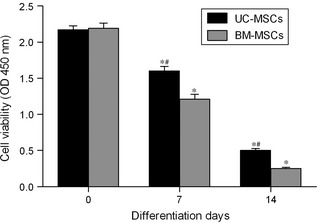
Analysis of UC ‐ MSC and BM ‐ MSC proliferation by CCK ‐8 assay. UC‐MSCs and BM‐MSCs were induced to differentiate into steroidogenic cells after being infected with adenovirus containing SF‐1 cDNA, and cultured for 7 and 14 days in the presence of cAMP. Control cultures were incubated growth medium alone (day 0). Values are means ± SD (n = 3). *P < 0.01 relative to respective controls. #P < 0.01 relative to differentiated BM‐MSCs.