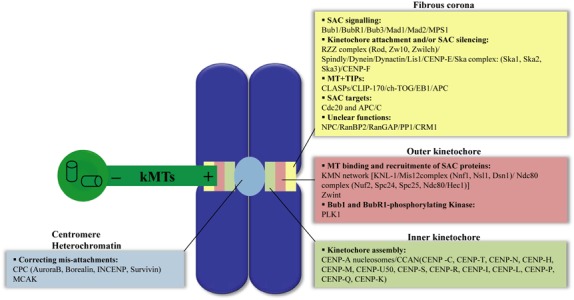
Figure 1.

Overview of protein complexes that build the kinetochore in animal cells. The kinetochore is built on the centromere as a trilaminar protein‐rich structure: the inner kinetochore, the outer kinetochore and the fibrous corona. Proteins that compose each kinetochore layer are grouped by function (APC/C, anaphase promoting complex/cyclosome; Bub1BubR1‐Bub3, budding uninhibited by benzimidazole; Cdc20, cell division cycle 20; CENP, centromere protein; CLASP, CLIP‐associating protein; CLIP170, cytoplasmic linker protein‐170; CPC, chromosome passenger complex; EB1, end‐binding protein‐1; INCENP, inner centromere protein; kMTs, kinetochore microtubules; LIS1, lissencephaly‐1; Mad1‐Mad2, mitotic‐arrest deficient; MCAK, mitotic centromere‐associated kinesin; MPS1, multipolar spindle‐1; MT, microtubules; NPC, nuclear pore complex; PLK1, polo‐like kinase‐1; RanBP2, Ran‐binding protein 2; RanGAP, Ran‐GTPase‐activating protein; RZZ, Rod (rough deal); SAC, spindle assembly checkpoint; Ska1–3, spindle and kinetochore‐associated proteins; Zw10, zeste white 10‐Zwilch complex; Zwint, Zw10 interactor. For details of dynamic localization of kinetochore proteins, see references (4, 92).
