
Figure 2.
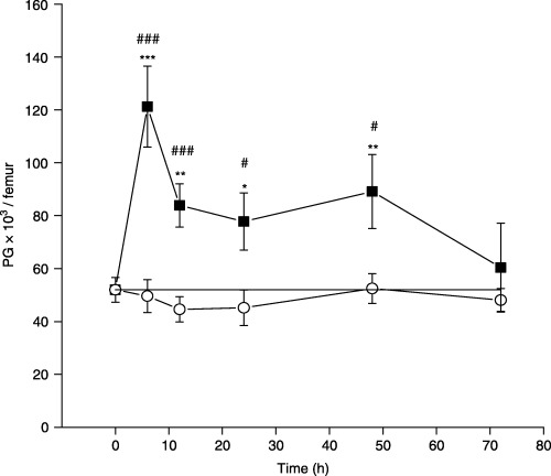
The effect of a single dose of rmIL‐17 on morphologically recognizable proliferative granulocytes (PG) in normal mice at different time points after treatment. The data points represent means ± SEM of five separate experiments. (–▪–) IL‐17 treated mice; (–○–) saline treated mice; (–—–) non‐treated mice. Significance at *P < 0.05, **P < 0.01, ***P < 0.001 for IL‐17‐ and saline‐treated vs. non‐treated mice, and at #P < 0.05, ###P < 0.001 for IL‐17‐treated vs. saline‐treated mice.