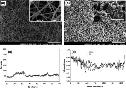
Figure 1.

Morphology of polyethersulphone ( PES ) (a) and bioactive glass ( BG )‐coated PES (b) magnifications (×1000; a, b) and with higher magnification (×12 000; a, b) (insets), XRD pattern of prepared BG nanoparticles: intensity of diffraction versus angle of radiation (2θ) (c), FTIR‐ATR spectra of pristine‐ and BG‐coated PES electrospun nanofibres (d).
