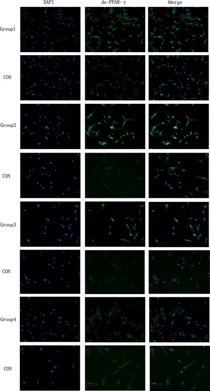
Figure 3.

Low‐intensity pulsed ultrasound ( LIPUS ) promoted de‐PPAR‐γ protein expression. After 2 + 5 days (Group 1), 2 + 3 days (Group 2), 5 days (Group 3), 3 days (Group 4) of adipogenic induction. Immunofluorescence staining was performed using anti‐de‐PPAR‐γ (green). Cell nuclei were counterstained with 4, 6‐diamidino‐2‐phenylindole (blue). (Magnification ×100).
