
Figure 2.
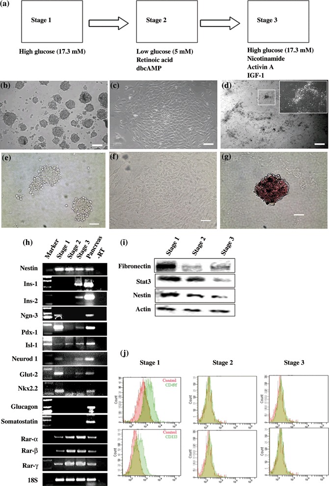
Developments of cells expressing islet markers from skin‐derived precursors ( SKPs). (a) Sketch of SKP differentiation protocol. (b–d) Stage‐specific cell cluster morphology; back arrow shows islet‐like cluster. (e–g) Dithizone staining of Stages 1–3 cells. Islet‐like clusters in Stage 3 were stained distinct crimson red by using dithizone (g). Cell in Stages 1 and 2 are not stained (e and f). (h). RT‐PCR analysis of gene expression during Stages 1–3 of pancreatic and endodermal markers. Pancreas RNA served as a positive control. (i) Western blot analysis of stem cell markers during Stage 1–3. (j) Examination of CD133 and CD49f, conserved marker of pancreatic progenitor, by flow cytometric analysis during Stage 1–3. Scale bar: 100 µm.