
Figure 3.
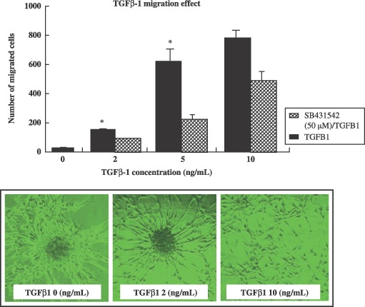
The effect of transforming growth factor‐β1 (TGF‐β1) on cultured neural progenitor cell migration. Cells were transferred to culture dishes containing serum‐free growth medium, which consisted of NB medium with B27 supplement and TGF‐β1 (10 ng/mL) with or without ALK inhibitor (SB431542) pre‐treatment. Cells were grown as neurospheres in Petri dishes, or in poly‐D‐lysine coated culture dishes. Cultured neurospheres, transferred to transwell membranes (Costar, 8 µm pore size) were coated on both sides with laminin, and were placed in 6‐well plates. Below the membrane, TGF‐β1 containing NB medium with B27 supplement was added to each well. To the upper chamber, NPC cells were incubated overnight in a 37 °C CO2 incubator. Cells on the lower surface were air‐dried and counterstained with Harris’ haematoxylin for 20 min, then washed. The stained inserts were placed on object slides, and numbers of cells on lower surfaces were assessed at ×200 using an inverted bright field microscope. Migration is expressed as a percentage (of cells per field) of spontaneous migration towards the cell bottom. Data are expressed as the means ± standard deviation (*P < 0.001).