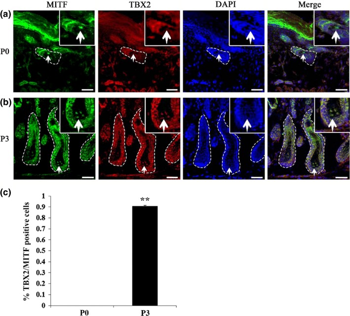
Figure 1.

TBX2 expressed in hair follicle melanocytes in mouse skin. (a, b) Double immunolabelling for MITF and TBX2 in postnatal hair follicles of mouse skin at P0 (a) and P3 (b). MITF staining specifically labelled melanocytes. While MITF expression (green nuclear staining) was detected in follicular melanocytes at P0, TBX2 expression (red nuclear staining) was only detected at P3. (c) Percentage of TBX2 and MITF double‐positive cells in hair follicle melanocytes of P0 and P3 mouse skins. Experiments were performed in triplicate and are represented as mean ± SD. Significance was determined by Student's t‐test whereby **P < 0.01. White arrows indicate positive cells. Dotted lines label the boundaries of hair follicles. Bar = 50 μm.
