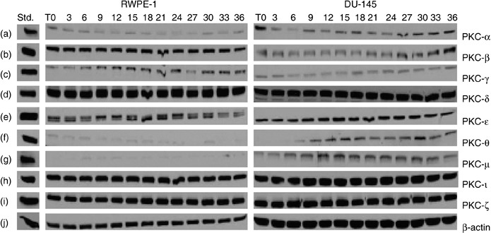
Figure 2.

Protein kinase C (PKC) isoforms in RWPE‐1 and DU‐145 cells. Cells were grown to 50–60% confluence. At this confluence, cells have space for cell‐cycle progession and cell division, without contact inhibition. Growth medium was removed and cells were semisynchronized by serum starvation for 48 h (T = 0). Growth media were then added to flasks and cells were allowed to grow for the indicated time (3–36 h). Flasks were collected every 3 h and cells were lysed as described in the Material and methods section. Equal amounts of protein (50–100 µg) were loaded on each lane and ran on 10% SDS‐PAGE. Column 1 contains standards for PKCs, column 2 the PKCs isozymes from normal RWPE‐1 cells, and column 3 PKCs isozymes in androgen‐independent prostate carcinoma DU‐145 cells at indicated times. Only nine PKC isozymes were present: PKC‐α, PKC‐βΙ, PKC‐γ, PKC‐δ, PKC‐ɛ, PKC‐θ, PKC‐µ, PKC‐ι and PKC‐ζ in both RWPE‐1 and DU‐145 cell lines (a–i). Higher protein concentration was required to detect PKC‐θ and PKC‐µ (150 µg each) and only traces were observed in normal RWPE‐1 cells (f–g). Regulation of PKC‐θ expression with the cell cycle was observed in DU‐145 cells (f). Western blots for PKC‐βΙΙ and PKC‐η were performed at higher concentration of protein (200 µg) for each cell line but neither enzyme was expressed. β‐actin (j) verified that equal amounts of protein were loaded in each lane.
