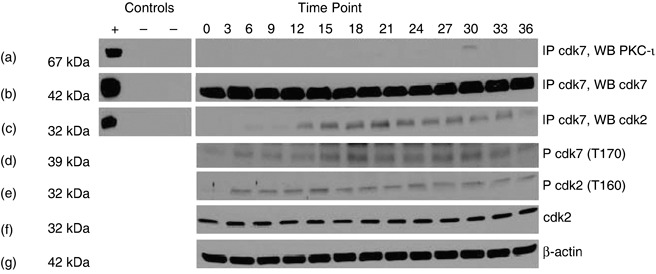
Figure 3.

Association of PKC‐ι, with Cdk7 in RWPE‐1 cells. Whole cell extracts (1 mg) from each time point were immunoprecipitated with rabbit polyclonal anti‐Cdk7 (5 µg) as described in the Materials and methods section. Column 1 contains both positive (+) and two negative controls (–). Positive control (+) is the whole cell lysates (100 µg); the first negative control (–) contains whole cell lysates (1 mg) plus rabbit IgG whole molecule (50 µl of 1 : 1 v/v) and the second negative control contains whole cell lysate (1 mg) plus rabbit IgG whole molecule (50 µl) and normal rabbit IgG serum (5 µg). Column 2 is cell lysates taken at indicated time points. Immunoprecipitates were separated by SDS‐PAGE and were Western blotted with anti‐PKC‐ι mouse monoclonal antibody. Physical association of PKC‐ι and Cdk7 were observed at 30 h time points (a). Immunoprecipitation (IP) with rabbit polyclonal Cdk7 (b) showed that Cdk2 is also coimmunoprecipitated (c). Western blotting of phospho‐Cdk7 (p‐Cdk7; T170) (d) and phospho‐Cdk2 (p‐Cdk2; T160) were also observed (e). Presence of Cdk2 was observed throughout the cell cycle (f) and β‐actin (g) shows equal loading of samples in each lane.
