Figure 5.

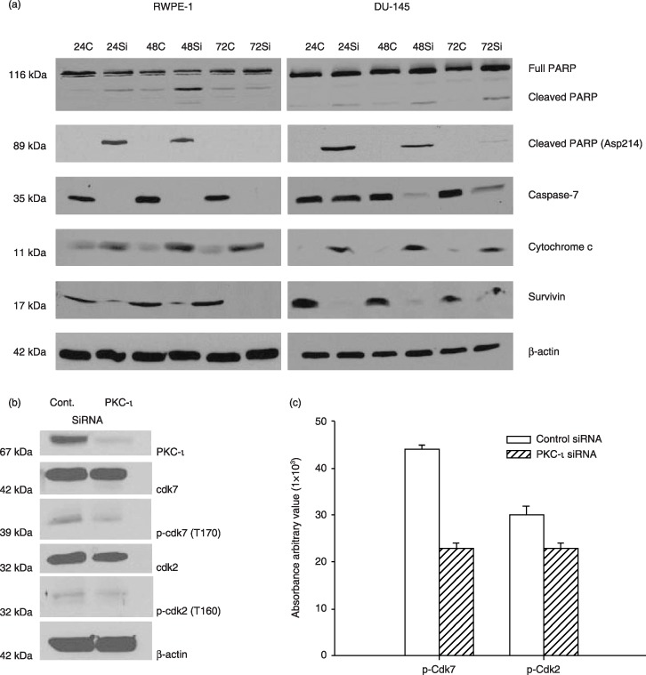
PKC‐ι siRNA leads to apoptosis. Whole cell extracts of RWPE‐1 treated with control siRNA and PKC‐ι siRNA were prepared as described in the Materials and methods section and immunoblot analysis of PARP and cleaved PARP (Asp214), indicated cells are undergoing apoptosis via activation of caspase‐7. Immunoblotting for survivin and cytochrome c further demonstrated apoptosis in PKC‐ι siRNA treated cells. Western blot analysis of β‐actin shows that an equal amount of protein was loaded in each lane. Similar immunoblots were performed for DU‐145 cells. Activation of PARP and caspase‐7, combined with an increase in cytochrome c and decrease in survivin indicated apoptosis in DU‐145 cells. (b) A separate experiment was repeated for RWPE‐1 cells with control siRNA and PKC‐ι siRNA and whole cells extracts (150 µg) were analysed for Cdk7, p‐Cdk7 (T170), Cdk2, p‐Cdk2 (T160), and β‐actin to verify equal loading of protein. (c) Densitometry for p‐Cdk7 and p‐Cdk2 showed a significant decrease in PKC‐ι siRNA‐treated cells compared to control siRNA (P = 0.001 and 0.035, respectively).
