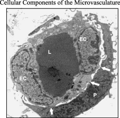
Figure 1.

Transmission electron micrograph of dermal microvessel showing the endothelial–pericyte functional unit. The pericyte (P) envelops a capillary lined with two endothelial cells (EC). Note the numerous cytoplasmic interdigitations and adhesion complexes between pericyte and endothelium (arrow). Magnification ×5600.
