
Figure 3.
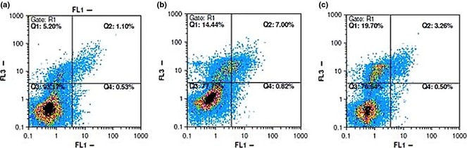
Flow cytometric analysis of annexin‐V/PI to quantify compound‐induced apoptosis in MCF ‐7 cells. (a) Dot plot of MCF‐7 cells with DMSO treatment for 48 h. (b) Dot plot of MCF‐7 cells with compound 6 treatment at 50.98 μm for 48 h. (c) Dot plot of MCF‐7 cells with compound 7 treatment at 21.30 μm for 48 h. Results shown are representative of three independent experiments. Quadrant 3, living cells An−/PI−; quadrant 4, early apoptotic cells An+/PI−; quadrant 2, late apoptotic cells An+/PI+; quadrant 1, necrotic cells An−/PI+.