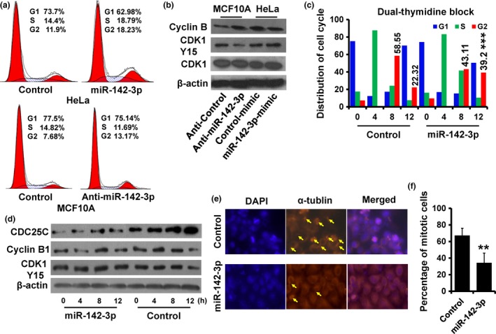
Figure 4.

Over‐expression of miR‐142‐3p lead to cell arrest at G2/M. (a) Cell cycle distribution of indicated cells was analyzed by flow cytometry analysis. (b) Expression of Cyclin B1, CDK1 and CDK1‐pY15 as detected by immunoblotting. (c) Cell cycle distribution of miR‐142‐3p‐overexpressed and control HeLa cells analyzed by flow cytometry. These cells were synchronized at the G1/S transition by a double‐thymidine block and released for the indicated times. (d) Expression of Cyclin B1, CDC25C and CDK1‐pY15 in cells as treated in (c) was detected by immunoblotting. (e), Immunofluorescence staining of DAPI (blue) and α‐tublin (orange) in cells sychronized as in (c) after 12 h release; arrow, mitotic cells. (f) Abundance of mitotic cells as in (e) was analyzed. **P < 0.01.
