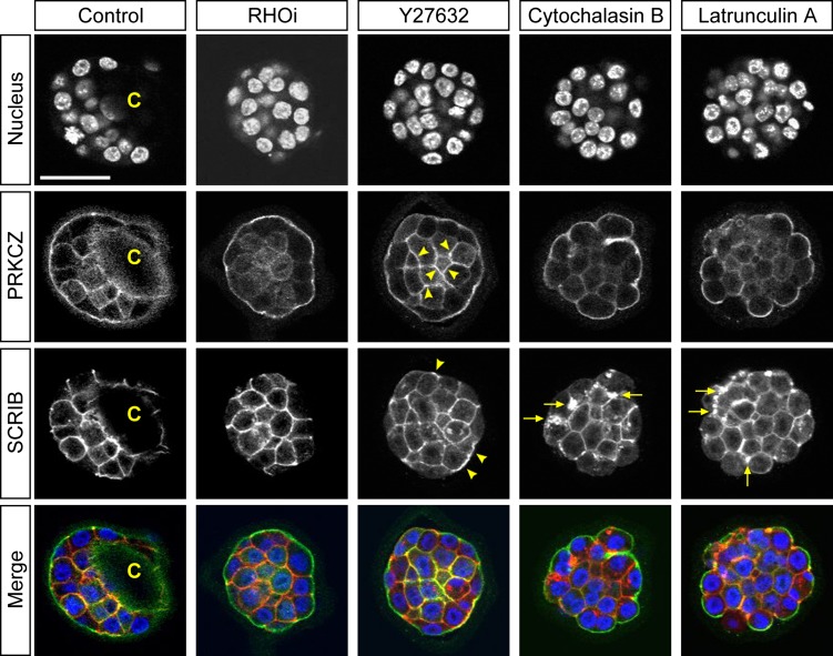
Figure 9.
Localization of cell polarity proteins in inhibitor-treated mouse blastocysts. Optical section confocal images of nucleus (blue), apical marker protein kinase C zeta (PRKCZ; green), and basolateral marker scribbled planar cell polarity (SCRIB; red) in representative E3.5 blastocysts that were treated for 8 h with no inhibitor (Control; n = 12), RHO inhibitor I (RHOi; n = 14), Y27632 (n = 14), cytochalasin B (n = 14) or latrunculin A (n = 14). C: Blastocyst cavity. Arrowheads indicate ectopic cortical enrichment for PRKCZ in inner cells and ectopic SCRIB localization to apical domain with Y27632 treatment. Arrows indicate aggregates of SCRIB in basolateral domain upon actin disruption. Scale bar = 50 μm.

