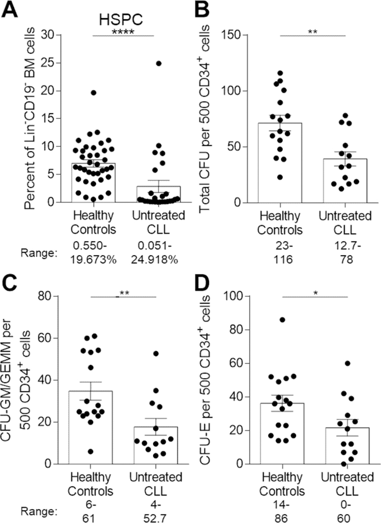
Figure 2. Reductions in BM HSPC frequency and functional clonogenic progenitors in CLL patients.

(A) Freshly isolated ACK-lysed BM cells were stained with combinations of antibodies to distinguish frequencies of Lin-CD19-CD34+ HSPCs between HCs (n=37) and CLL patients (n=26) (57 individual experiments). Data is represented as the mean and SEM with each point indicating an individual BM sample. Numbers under the figure represent the range for each cohort evaluated. (B-D) Numbers of (B) total CFU, (C) CFU-granulocyte, monocyte/granulocyte, erythrocyte, monocyte, megakaryocyte (CFU-GM/GEMM), or (D) CFU-erythroid (CFU-E) colonies per 500 input CD34+ cells. HC n=16 and CLL n=13 (24 individual experiments). Data is represented as the mean and SEM of triplicate platings with each point indicating an individual subject. Numbers under each figure represent the range for each colony evaluated for HCs and CLL patients *P<0.05, **P<0.01, and ****P<0.0001 were obtained by Mann-Whitney U-test.
