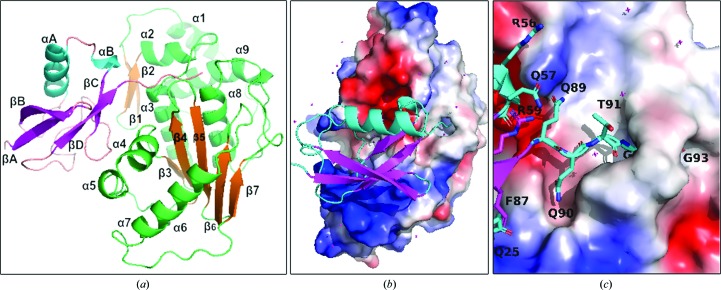
Figure 2.
The overall structure, domain definition and electrostatic map of the SENP1–SUMO2 structure. (a) Secondary-structural elements of SENP1 (shown in green/orange) and SUMO2 (shown in cyan/magenta), which are named consecutively starting from the N-terminus. (b) The general orientation of SUMO2 on SENP1 showing the parts of that occupy electropositive (blue) and electronegative (red) regions of SENP1. (c) The QQTGG motif of SUMO2, which is deeply buried in the subpocket of the active site of SENP1 to provide the predominant interaction for the recognition.

