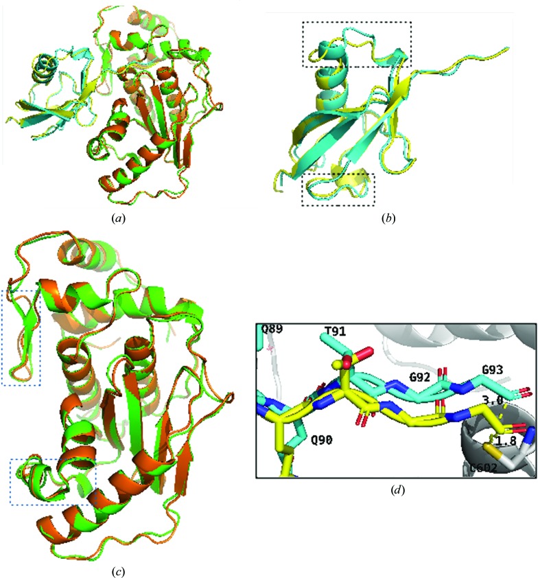
Figure 4.
Comparison of the structures of SENP1 and SUMO2 with the published covalent structure. (a) Overall alignment of the SENP1–SUMO2 complex with the reference stucture (Shen, Dong et al., 2006 ▸). (b) Alignment of SUMO2. (c) Alignment of SENP1. The current structure is shown in green (SENP1) and cyan (SUMO2) and the reference structures in yellow (SUMO2) and orange (SENP1). The alignment shows that major differences, shown in boxes, arise from the SENP1–SUMO2 interaction interface. (d) Alignment of residues of the QQTGG motif and the distance between Cys602 of SENP1 (gray) and the Gly93 residue of SUMO2.

