Figure 1.
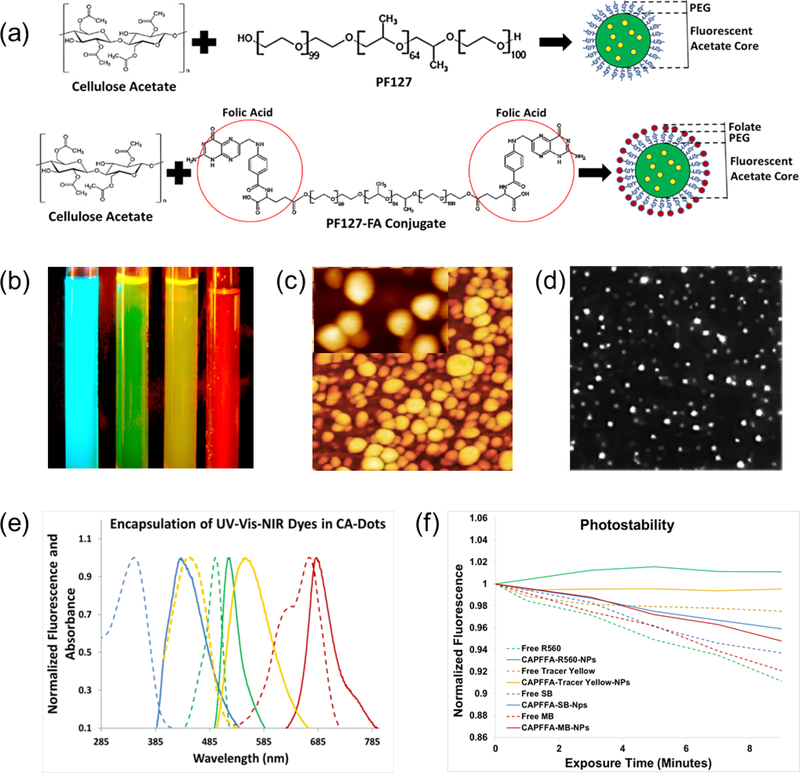
Assembly of CA-dots and their basic properties. (a) Schematics of the assembly. A physical mix of cellulose acetate dissolved in an organic solvent and pluronic acid F127 precipitate in an aqueous phase to produce PEG-coated CA-dots; folate functionalized CA-dots are assembled in a similar way by substituting F127 “module” with FA-F127 conjugate. (b) Fluorescent image of four tubes with CA-dots containing encapsulated Stilbene 420 (blue), Rhodamine 560 (green), Tracer Yellow (yellow), and Methylene Blue (red/NIR); fluorescence of blue, green, and yellow CA-dots were excited with UV flashlight, and the NIR dye with 660nm red laser. The image was taken with Fuji Pro camera with extended NIR sensitivity. (c) 1×1 μm2 AFM image of CA nanoparticle (CA-SB-PEG-FA); insert is 170×270nm2 image showing about spherical geometry of the particles; (d) 25×25 μm2 fluorescent image of single CA-dots near the surface of a glass slide; (e) absorbance and fluorescence spectra of CA-dots with encapsulated different fluorescent dyes: blue (ex/em: 365 nm/420 nm; Stilbene 420), green (ex/em: 495 nm /530 nm; Rhodamine 560), yellow (ex/em: 450 nm /560 nm; Tracer Yellow), and red/NIR (ex/em: 665 nm/680 nm; Methylene Blue) maxima of fluorescent intensity. (e) photostability of the obtained particles and the corresponding free dye; a short-time exposure is shown in which photodegradation kinetics are comparable (relative beaching <10%).

