Figure 2.

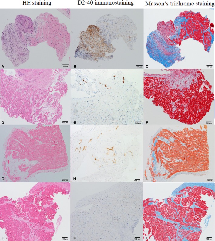
Representative cases of histological cardiac sarcoidosis (A through C), clinical cardiac sarcoidosis (CS; D through I), and idiopathic dilated cardiomyopathy (DCM) (J through L). A, D, G, and J, Hematoxylin‐eosin (HE) staining ×100 original magnification. B, E, H, and K, D2‐40 immunostaining ×100 original magnification. C, F, I, and L, Masson trichrome staining ×100 original magnification. In histological CS, routine HE staining (A) shows noncaseous granuloma consisting of giant cells, epithelioid cells, and fibroblasts. With D2‐40 immunostaining (B), numerous numbers of small lymphatic capillaries were elucidated within granuloma. Endomyocardial granuloma with fibrosis is shown by Masson trichrome staining (C). In clinical CS, no granuloma is seen in standard HE staining (D and G). There is an increased number of lymphatic vessels within connective tissues of fibrosis area (E and H). Fibrosis tends to be mosaic pattern with relatively preserved myocardium (F and I). In DCM, there are extensive myocardial damages seen in HE staining (J). No lymph duct or few sporadic lymph ducts are observed (K). Replacement fibrosis is seen in a diffuse or focal pattern of myocardium that is extensively damaged (L).
