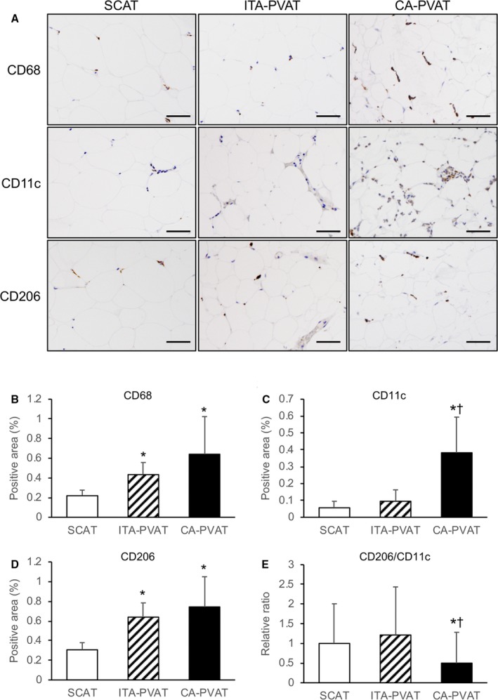
Figure 3.

Macrophage infiltration in fat pads. A, Representative immunohistological staining, including CD68 (a marker of macrophages), CD11c (a marker of M1 macrophages), and CD206 (a marker of M2 macrophages), of subcutaneous adipose tissue (SCAT) and of perivascular adipose tissue surrounding the internal thoracic artery (ITA‐PVAT) and that surrounding the coronary artery (CA‐PVAT). Bar=50 μm. B through D, Positive areas of immunohistological staining of CD68, CD11c, and CD206 in SCAT, ITA‐PVAT, and CA‐PVAT (n=10 in each group). E, Ratios of the macrophage infiltration areas of CD206 to CD11c in SCAT, ITA‐PVAT, and CA‐PVAT. Results are shown as mean±SD. *P<0.05 vs SCAT; † P<0.05 vs ITA‐PVAT.
