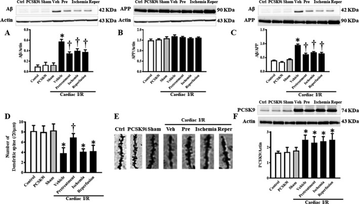
Figure 4.

Effects of PCSK9 inhibition on Aβ and APP protein expression levels and dendritic spine density after cardiac I/R injury. A, Protein levels of Aβ established by Western blotting, normalized to actin. B, Protein levels of APP by Western blotting, normalized to actin. C, Protein levels of Aβ by Western blotting, normalized to APP. D, Dendritic spine density per 20 μm apical tertiary dendrite; E, Representative images of dendritic spines. F, PCSK9 levels by Western blotting, normalized to actin. Aβ, amyloid beta; I/R, ischemia/reperfusion; Veh, vehicle; Pre, pretreatment; Reper, reperfusion; PCSK9, Proprotein convertase subtilisin/kexin type 9; PCSK9i, proprotein convertase subtilisin/kexin type 9 inhibitor; Control, rat group with no surgical intervention; PCSK9i, rats group with no intervention and treated with PCSK9i for 180 minutes; Sham, rat group with no surgical intervention/treatment; Vehicle, rats treated with vehicle at 15 minutes before cardiac I/R; Pretreatment, rats treated with PCSK9i at 15 minutes before cardiac I/R; Ischemia, rats treated with PCSK9i at 15 minutes during ischemic period; Reperfusion, rats treated with PCSK9i at the onset of reperfusion period (n=4–10 per group). *P<0.05 vs sham, † P<0.05 vs vehicle.
