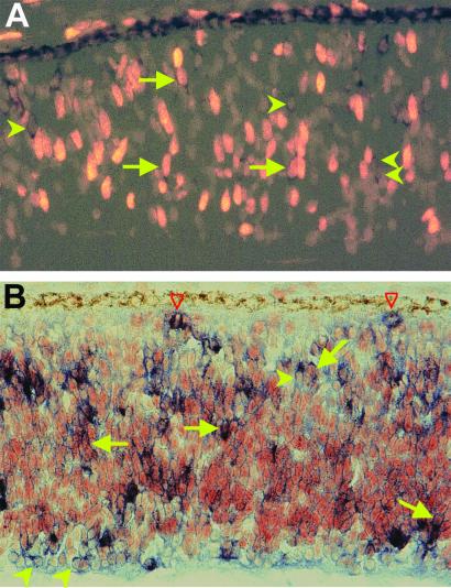
Figure 1.
Expression of ngn2 in proliferating cells of E7 chick retinal neuroepithelium. (A) Doubling-labeling for ngn2 mRNA (dark stains in the cytoplasm) and BrdUrd incorporation (bright nuclei). (B) Doubling-labeling for ngn2 mRNA (blue stains in the cytoplasm) and PCNA immunoreactivity (red/brown stains of the nuclei). Arrows point to double-labeled cells. Arrowheads point to ngn2+/BrdUrd− or ngn2+/PCNA− cells. Red open arrowheads point to ngn2+ cells that were in M-phase of the cell cycle. Magnification: ×100.

