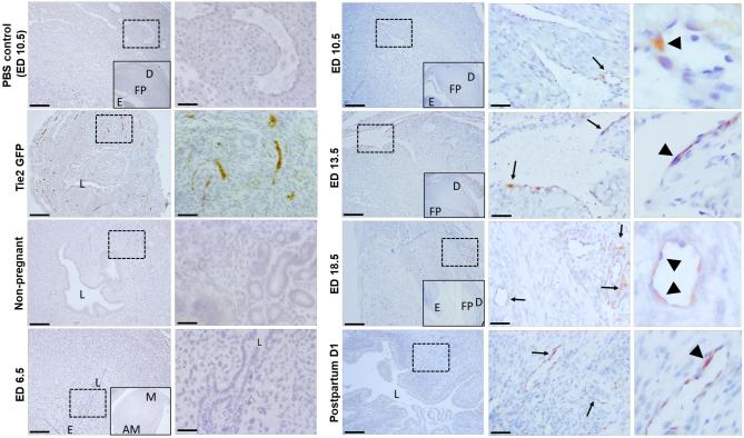
Figure 4.
Immunohistochemical detection of GFP-positive endothelial cells in uterine sections. Immunostaining of uterine sections using anti-GFP antibody. The inset within left panels of pregnant sections (ED6.5, ED10.5, ED13.5, ED18.5, and PBS ED10.5 control) is a lower magnification image of the panel for orientation, showing the mesometrial (M) and anti-mesometrial (AM) areas, the decidua (D), fetal side of the placenta (FP) and embryo (E). The right panels of each time point are higher magnification images of the dashed respective rectangle area in the left panel. GFP-positive endothelial cells (brown) are seen in uterine blood vessels of Tie2-GFP positive control mice while no GFP positive staining is seen in PBS, non-pregnant and ED6.5 groups. Arrows point to decidual blood vessels with incorporated GFP-positive endothelial cells (arrowheads) in ED10.5, ED13.5, ED18.5, and postpartum day 1 groups. Representative of 3–4 mice for each group. Scale bars in the lower magnification images = 200 um. Scale bars in the higher magnification images = 50 um.

