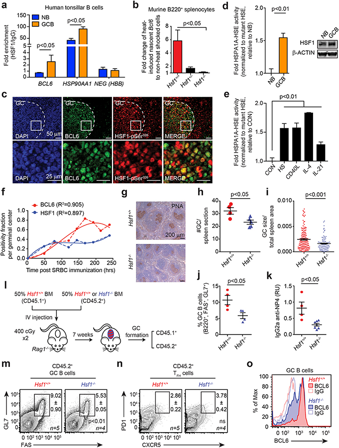
Figure 2. B-cells require HSF1-dependent BCL6 induction for optimal germinal center formation.
a, Enrichment of HSF1 in NB and GC B-cells at the BCL6 promoter, HSP90AA1 promoter and a negative control HBB (representative of 3 biological replicates). b, Nascent Bcl6 mRNA in heat shocked murine B220+ splenocytes of Hsf1+/+, Hsf1+/− and Hsf1−/− mice normalized to Hprt1 (n=3 mice per genotype). c, Immunofluorescence of paraffin-embedded serial human tonsillar sections. d, AlphaLisa activity of HSPA1A HSE from nuclear protein of human tonsillar NB and GC B-cells (n=5 pooled replicates) with accompanying immunoblot for total HSF1 (right). e, AlphaLisa activity of the consensus HSPA1A HSE from nuclear protein from human splenic NBs resting (CON), heat-shocked (HS) or treated with immune stimuli (n=4–7 pooled replicates). f, Fraction of PNA+ HSF1+ (blue) or PNA+ BCL6+ (red) cells per GC post-immunization as a function of time. Polynomial fits (solid line) of real data points (dashed line) are shown. g-j, Representative PNA staining (g), GC size (h), GC number (i) and percentage (j) of Hsf1+/+ and Hsf1−/− mice after immunization (n=4 mice per genotype). k, Titers of high-affinity NP-specific IgG2a from Hsf1+/+ and Hsf1−/− mice after immunization with NP-CGG (mean ± s.e.m., n=4–6 mice per genotype). l-n, Bone marrow chimeras generated with CD45.1+ Hsf1+/+ and CD45.2+ Hsf1+/+ or Hsf1−/− mice (l). Representative flow cytometry plot of CD45.2+ GC B-cells (m) and GC TFH cells (n) from mice after immunization (n=4–5 mice per genotype). See Supplementary Fig. 3c–e for chimerism and CD45.1+ GC B-cells and GC TFH cells. o, BCL6 staining intensity in splenic GC B-cells from Hsf1+/+ and Hsf1−/− mice after immunization. P values were calculated by two-sided T-test. Data presented as mean ± s.e.m.

